Understanding the epidemiology, diagnosis, and management of AL Amyloidosis. Excellent review article @JACCJournals for students and residents. Thanks for the recommendation @daehyunleemd @USFIMres
#cardiology #cardiotwitter #amyloidosis #MedTwitter
#cardiology #cardiotwitter #amyloidosis #MedTwitter
Background:
- AL Amyloidosis increasingly diagnosed 2/2 to misfolded proteins (monoclonal immunoglobulin light chains) in 1 or more body tissues
- Can be localized/systemic, benign/life-threatening
- Most common forms are ATTR (transthyretin) or AL (light-chain) amyloidosis
- AL Amyloidosis increasingly diagnosed 2/2 to misfolded proteins (monoclonal immunoglobulin light chains) in 1 or more body tissues
- Can be localized/systemic, benign/life-threatening
- Most common forms are ATTR (transthyretin) or AL (light-chain) amyloidosis
Pathology:
- "L" in AL amyloidosis is dominant protein depositing in AL amyloid fibrils
- Clonal light chains circulate in bloodstream and can deposit in any organ
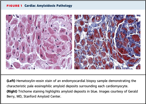
- In the heart, deposition found throughout myocardium, w/ amyloid deposits surrounding each cardiomyocyte
- "L" in AL amyloidosis is dominant protein depositing in AL amyloid fibrils
- Clonal light chains circulate in bloodstream and can deposit in any organ
- In the heart, deposition found throughout myocardium, w/ amyloid deposits surrounding each cardiomyocyte
Pathology:
- Increased wall thickness 2/2 amyloid fibril deposition leads to small end-diastolic volume, low stroke volume, decreased cardiac output
- Worse prognosis w/ AL > ATTR despite higher cumulative fibril deposition in ATTR
- Goal: lower circulating light chains
- Increased wall thickness 2/2 amyloid fibril deposition leads to small end-diastolic volume, low stroke volume, decreased cardiac output
- Worse prognosis w/ AL > ATTR despite higher cumulative fibril deposition in ATTR
- Goal: lower circulating light chains
Light-Chain Production:
- Occurs w/ proliferation of a plasma cell clone in bone marrow. Also by:
- Clonal B-lymphocytes in lymphoma
- Clonal plasma cells outside bone marrow (plasmacytoma)
- Clonal plasma cells/B-lymphocytes within tissue in inflammation
- Occurs w/ proliferation of a plasma cell clone in bone marrow. Also by:
- Clonal B-lymphocytes in lymphoma
- Clonal plasma cells outside bone marrow (plasmacytoma)
- Clonal plasma cells/B-lymphocytes within tissue in inflammation
Diagnosis:
- Can present as localized (found incidentally on biopsy for different reason, i.e. gastritis on EGD with AL amyloid deposits) or systemic
- Traditionally CRAB (hypercalcemia, renal insufficiency, anemia, lytic bone lesions)
- Can present as localized (found incidentally on biopsy for different reason, i.e. gastritis on EGD with AL amyloid deposits) or systemic
- Traditionally CRAB (hypercalcemia, renal insufficiency, anemia, lytic bone lesions)
Diagnosis:
- New criteria: serum free light chain (FLC) ratio: involved/not-involved ≥ 100, clonal bone marrow plasma cells ≥ 60%, lytic detection on CT/MRI/PET
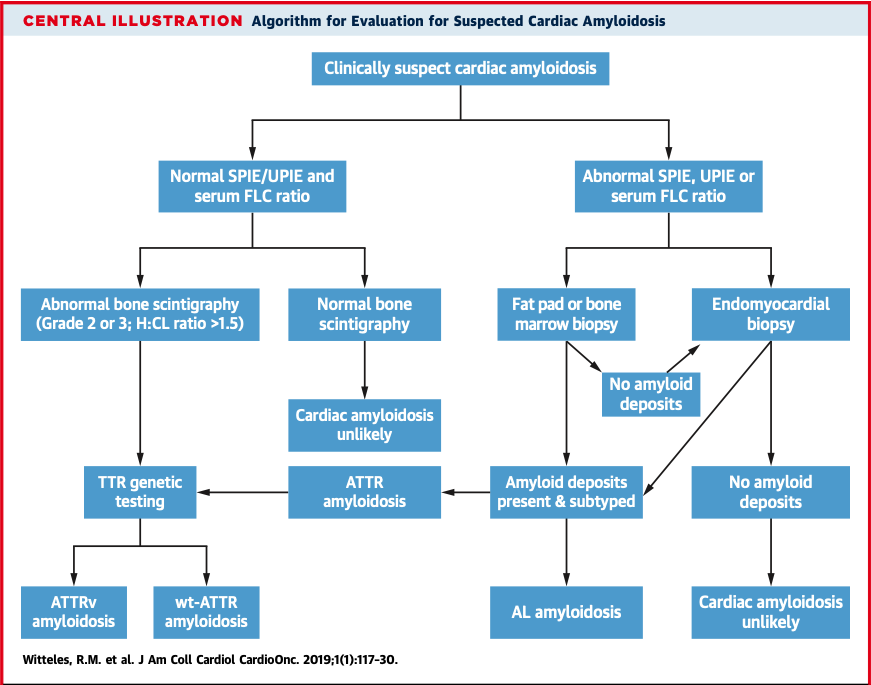
- Normal SPEP, UPEP, and serum FLC nearly rules out systemic AL amyloidosis
- Biopsy followed by mass spectometry
- New criteria: serum free light chain (FLC) ratio: involved/not-involved ≥ 100, clonal bone marrow plasma cells ≥ 60%, lytic detection on CT/MRI/PET
- Normal SPEP, UPEP, and serum FLC nearly rules out systemic AL amyloidosis
- Biopsy followed by mass spectometry
Cardiac Amyloid Clues:
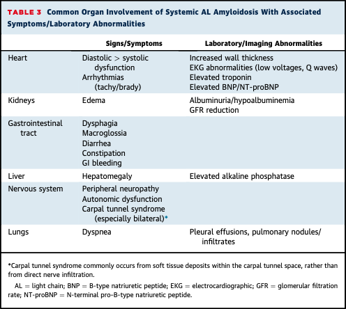
- Persistently elevated troponin (despite correlating cath) - HF w/ other organ findings (macroglossia, nephrotic syndrome, elevated alkaline phosphatase, dysphagia)
- Heart block, atrial arrhythmias, ventricular arrhythmias w/ increased thick ventricle
- Persistently elevated troponin (despite correlating cath) - HF w/ other organ findings (macroglossia, nephrotic syndrome, elevated alkaline phosphatase, dysphagia)
- Heart block, atrial arrhythmias, ventricular arrhythmias w/ increased thick ventricle
Imaging:
- ECHO: increased wall mass in LV & RV in concentric/diffuse pattern, thickening inter-atrial septum/valves, pericardial effusion
- EKG: normal voltage in setting of ventricular hypertrophy should be red flag
- Cardiac MRI: Diffuse non-ischemic gadolinium uptake
- ECHO: increased wall mass in LV & RV in concentric/diffuse pattern, thickening inter-atrial septum/valves, pericardial effusion
- EKG: normal voltage in setting of ventricular hypertrophy should be red flag
- Cardiac MRI: Diffuse non-ischemic gadolinium uptake
Treatment:
- Light chain suppressive therapy with alkylator (Melphalan) + steroid (prednisone) was first regimen
- Autologous Stem Cell transplantation new option, but tight selection criteria
- CyBorD (cyclophosphamide, bortezomib, and dexamethasone) w/ good responses
- Light chain suppressive therapy with alkylator (Melphalan) + steroid (prednisone) was first regimen
- Autologous Stem Cell transplantation new option, but tight selection criteria
- CyBorD (cyclophosphamide, bortezomib, and dexamethasone) w/ good responses
Treatment:
- Most promising recent agent is Daratumumab, antibody-directed against CD38 (cell surface protein on clonal plasma cells): study w/ 25 patients who were refractory to multiple agents had 76% response rate
- Most promising recent agent is Daratumumab, antibody-directed against CD38 (cell surface protein on clonal plasma cells): study w/ 25 patients who were refractory to multiple agents had 76% response rate
Cardiac Management:
- Standard HF often poorly tolerated (B-blockers, ACE/ARB) can worsen bradycardia/heart block
- Anticoagulation b/c high risk for thrombus formation
- ICD placement w/ >1 yr life expectancy w/ evidence of ventricular arrhythmias
- Cardiac transplant
- Standard HF often poorly tolerated (B-blockers, ACE/ARB) can worsen bradycardia/heart block
- Anticoagulation b/c high risk for thrombus formation
- ICD placement w/ >1 yr life expectancy w/ evidence of ventricular arrhythmias
- Cardiac transplant
Conclusion:
- AL amyloidosis w/ cardiac manifestations requires high degrees of suspicion
- Need collaboration w/ hematologists, cardiologists, and other sub-specialists
- Disease remains life-threatening, but advances in chemotherapy and immunotherapy
- Early dx is critical
- AL amyloidosis w/ cardiac manifestations requires high degrees of suspicion
- Need collaboration w/ hematologists, cardiologists, and other sub-specialists
- Disease remains life-threatening, but advances in chemotherapy and immunotherapy
- Early dx is critical

 Read on Twitter


