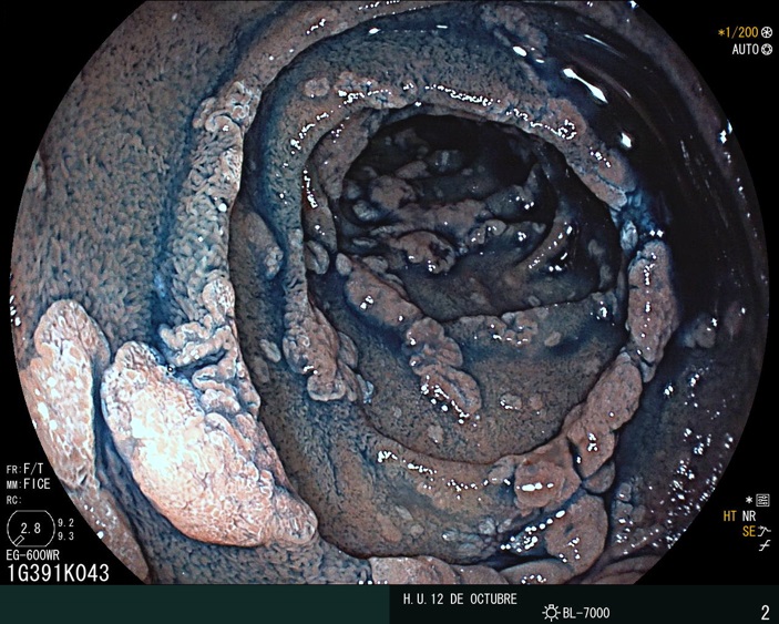
Duodenal adenomas (picture) are a common problem in the upper GI of the patients with familial adenomatous polyposis (FAP)
However, adenomas and gastric cancer are a matter of increasing concern
A thread with a poll at the end
@SEEDendoscopia @aegastro
However, adenomas and gastric cancer are a matter of increasing concern
A thread
 with a poll at the end
@SEEDendoscopia @aegastro
With regard to gastric cancer, most cases arise in patients with a carpeting of fundic gland polyposis and associated with large mounds of proximal gastric polyps.
In this example, capsule endoscopy detected only small fundic gland polyps
In this example, capsule endoscopy detected only small fundic gland polyps
Data regarding gastric adenomas are sparse and the existence of a gastric-cancer genotype-phenotype correlation was not obvious in the adenoma group in this study led by Dr. @AndyLatchford https://doi.org/10.1007/s10689-017-9966-0
And, how can we manage gastric adenomas in these patients?
Recently, an observational study by combining data from St. Mark’s and Amsterdam UMC have been published.
Gastric adenomas were observed in 14% of patients and endoscopic resection was safe
https://www.thieme-connect.de/products/ejournals/abstract/10.1055/a-1265-2716
Recently, an observational study by combining data from St. Mark’s and Amsterdam UMC have been published.
Gastric adenomas were observed in 14% of patients and endoscopic resection was safe
https://www.thieme-connect.de/products/ejournals/abstract/10.1055/a-1265-2716
Gastric adenomas > 20mm had a 33% risk of harboring HGD compared with 4 % in adenomas ≤ 20mm
ESD was performed in 2 cases of lesions > 20 mm and LGD was observed in both.
ESD was performed in 2 cases of lesions > 20 mm and LGD was observed in both.
ESD for these big lesions is indeed a useful endoscopic technique after optical diagnosis with magnification is done.
In LGD, regular V and S patterns are present. Regular WOS may also be seen as was the case with this patient with a pathogenic mutation in APC
In LGD, regular V and S patterns are present. Regular WOS may also be seen as was the case with this patient with a pathogenic mutation in APC
After a successful en bloc resection, the final specimen (37×37mm, tumor size 21×21mm) was retrieved. LGD was confirmed for this 0-IIa+IIc lesion (as observed in previous biopsies)
But another small lesion was also observed during the procedure
Chromoendoscopy and magnification were done
Chromoendoscopy and magnification were done

After watching this latter video, what is your diagnosis?

 Read on Twitter


