1/
Let's talk #cardiacamyloidosis (CA)— in fact, let's talk imaging in CA, but first things first: what is the gold standard for dx’ing CA?
Let's talk #cardiacamyloidosis (CA)— in fact, let's talk imaging in CA, but first things first: what is the gold standard for dx’ing CA?
2/
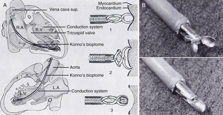
Congo staining of amyloid deposits in tissue obtained via endomyocardial biopsy (EMB) is the definitive way of dx'ing CA


EMB however is not widely available & pretty invasive. out the *OG* myotome from Konno et al in their 1963 article
https://pubmed.ncbi.nlm.nih.gov/14072735/
Congo
 staining of amyloid deposits in tissue obtained via endomyocardial biopsy (EMB) is the definitive way of dx'ing CA


EMB however is not widely available & pretty invasive.
 out the *OG* myotome from Konno et al in their 1963 article
https://pubmed.ncbi.nlm.nih.gov/14072735/
3/
Now let's consider imaging for CA:
- Widely available
- Noninvasive
- Quantitative
- Whole heart imaging can estimate amyloid burden
- Repeated w/ease to assess response to therapy
https://pubmed.ncbi.nlm.nih.gov/31607664/
Now let's consider imaging for CA:
- Widely available

- Noninvasive

- Quantitative

- Whole heart imaging can estimate
 amyloid burden
- Repeated w/ease to assess response to therapy

https://pubmed.ncbi.nlm.nih.gov/31607664/
4/
There are different ways to look at amyloid in the , each with their
  &
& , and own ways of
 amyloid— here we will focus on the two most common modalities:
1. Scintigraphy/SPECT (with -avid radiotracers like PYP)
2. Cardiac MRI
There are different ways to look at amyloid in the
 , each with their
  &
& , and own ways of
 amyloid— here we will focus on the two most common modalities:
 1. Scintigraphy/SPECT (with
 -avid radiotracers like PYP)
2. Cardiac MRI

5/
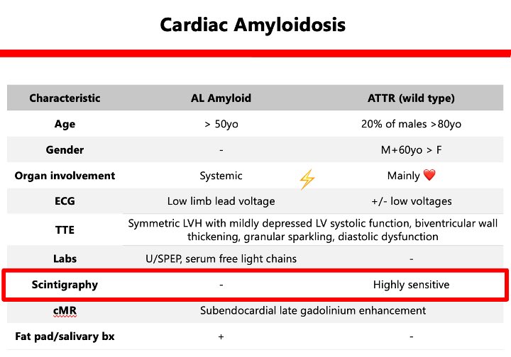
AL amyloid and ATTR (wild type) are the 2 most common types of #cardiacamyloid we encounter, so let's focus on them below and talk *scintigraphy/SPECT* first (check out our #TheZollCenter S2E2 for a deeper dive into the chart below )
)
AL amyloid and ATTR (wild type) are the 2 most common types of #cardiacamyloid we encounter, so let's focus on them below and talk *scintigraphy/SPECT* first (check out our #TheZollCenter S2E2 for a deeper dive into the chart below
 )
)
6/
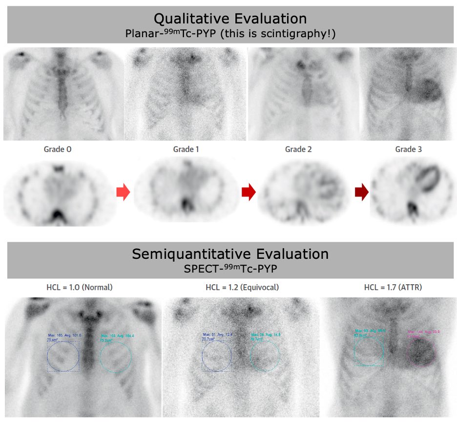
#Scintigraphy detects radiotracer displays 2D image (~XR)
#SPECT is similar, BUT contiguous 2D images (~CT)
- -avid markers like 99mTc-PYP are taken up by ATTR
- What we're unsure of: mechanism of uptake
- How to eval? grades or semiquant
https://pubmed.ncbi.nlm.nih.gov/31607664/
#Scintigraphy detects radiotracer
 displays 2D image (~XR)
#SPECT is similar, BUT
 contiguous 2D images (~CT)
-
 -avid markers like 99mTc-PYP are taken up by ATTR
- What we're unsure of: mechanism of uptake

- How to eval?
 grades or semiquant
https://pubmed.ncbi.nlm.nih.gov/31607664/
7/
How #sensitive is scintigraphy for ATTR CA? Incredibly
In a 2016 study by Gillmore et al., 857 pts w/histo-proven amyloid (374 w/EMB), scint was >99% sn & 86% sp for ATTR CA (false results were almost all from uptake in pts w/cardiac AL amyloidosis)
https://pubmed.ncbi.nlm.nih.gov/27143678/
How #sensitive is scintigraphy for ATTR CA? Incredibly
In a 2016 study by Gillmore et al., 857 pts w/histo-proven amyloid (374 w/EMB), scint was >99% sn & 86% sp for ATTR CA (false
 results were almost all from uptake in pts w/cardiac AL amyloidosis)
https://pubmed.ncbi.nlm.nih.gov/27143678/
8/
And lastly this out (are you sitting down?): Gillmore et al. also found that:
1: grades 2 or 3 myocardial tracer uptake
PLUS
2: absence of a monoclonal protein in serum or urine
EQUALS
 : Specificity and PPV for ATTR CA of 100%
And lastly
 this out (are you sitting down?): Gillmore et al. also found that:
 1: grades 2 or 3 myocardial tracer uptake
PLUS
2: absence of a monoclonal protein in serum or urine
EQUALS
 : Specificity and PPV for ATTR CA of 100%
9/
In fact, ATTR CA can be dx’ed at places w/limited access to EMB and in pts who decline or who are not candidates for invasive procedures—this is a #gamechanger
For #funsies: can you guess the grade in the images below?
In fact, ATTR CA can be dx’ed at places w/limited access to EMB and in pts who decline or who are not candidates for invasive procedures—this is a #gamechanger
For #funsies: can you guess the grade in the images below?
10/
The above was read as grade with semiquantitative eval giving a heart to contralateral lung ratio of 2.1— pt ultimately dx’ed w/ATTR CA
Now the following are the images of the case discussed on #TheZollCenter S2E2:
The above was read as grade
 with semiquantitative eval giving a heart to contralateral lung ratio of 2.1— pt ultimately dx’ed w/ATTR CA
Now the following are the images of the case discussed on #TheZollCenter S2E2:
11/
If you didn’t see much, then you’re , this was called grade
 w/no significant evidence of myocardial radiotracer uptake..
Quick #sidenote: keep in mind that -avid radiotracer images lack structural/hemodynamic info, so typically they are used alongside echo or CMR
If you didn’t see much, then you’re
 , this was called grade
 w/no significant evidence of myocardial radiotracer uptake..
 Quick #sidenote: keep in mind that
 -avid radiotracer images lack structural/hemodynamic info, so typically they are used alongside echo or CMR
12/
That’s all for now folks, stay tuned for the CMR portion of imaging in #cardiacamyloidosis this later this week! Thank you for joining!
And special thanks to Dorbala et al. for the incredible CA imaging review cited throughout https://pubmed.ncbi.nlm.nih.gov/31607664/
That’s all for now folks, stay tuned for the CMR portion of imaging in #cardiacamyloidosis this later this week! Thank you for joining!
And special thanks to Dorbala et al. for the incredible CA imaging review cited throughout https://pubmed.ncbi.nlm.nih.gov/31607664/

 Read on Twitter


