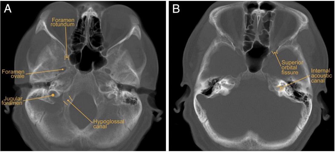
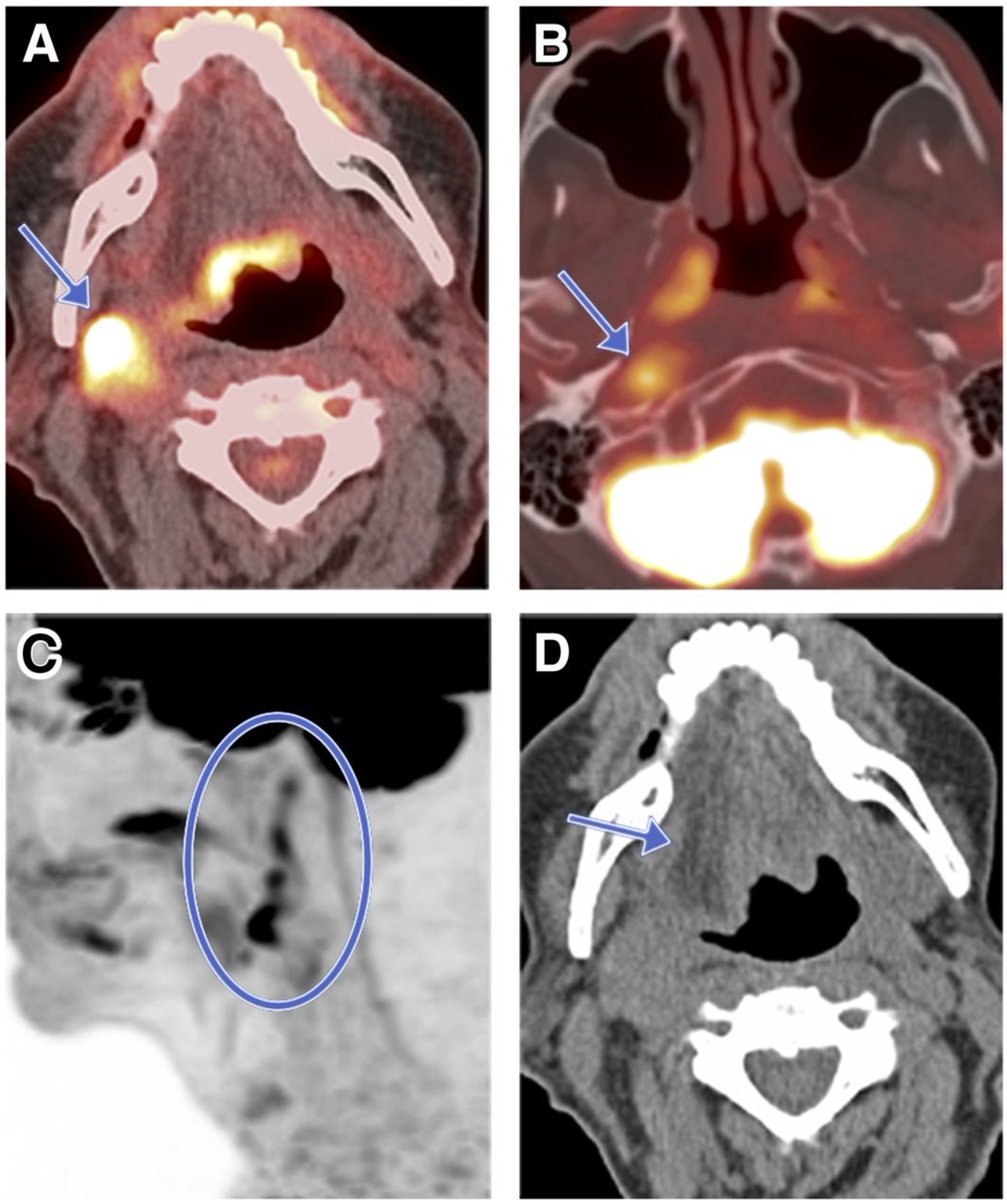
The most common nerves involved in perineural tumor spread in the head are CN VII and V, followed by CN XII.
In cancer cases, inspect at their courses and skull base foramina.
https://jnm.snmjournals.org/content/60/3/304/tab-figures-data
https://jnm.snmjournals.org/content/jnumed/suppl/2019/03/01/jnumed.118.214312.DC1/214312_Supplemental_Data.pdf
In cancer cases, inspect at their courses and skull base foramina.
https://jnm.snmjournals.org/content/60/3/304/tab-figures-data
https://jnm.snmjournals.org/content/jnumed/suppl/2019/03/01/jnumed.118.214312.DC1/214312_Supplemental_Data.pdf
Go through a mental checklist for cases of cancer on the head: FAT PADS. FAT PADS. FAT PADS.
https://insightsimaging.springeropen.com/articles/10.1007/s13244-018-0672-8
https://insightsimaging.springeropen.com/articles/10.1007/s13244-018-0672-8
The pre-contrast T1 WITHOUT fat suppression is a crucial part of head and neck MRI.
Look for the clean fat pads, which you don't get with fat-sat due to susceptibility artifacts
http://doi.org/10.3174/ng.9170238
Look for the clean fat pads, which you don't get with fat-sat due to susceptibility artifacts
http://doi.org/10.3174/ng.9170238
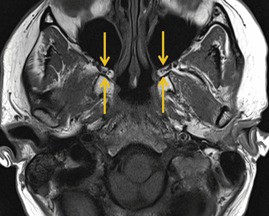
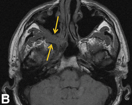
The pterygopalatine fossa is the key stop for perineural tumor spread coming along CN V2 branches from the orbit, sinonasal tract, and palate.
Normal case vs. evil grey in the right PPF:
Normal case vs. evil grey in the right PPF:
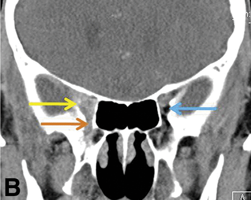
The orbital apex has fat. Tumor squeezing along cranial nerves through the superior and inferior orbital fissures and optic canal will blur this fat.
Melanoma on the right vs. normal on the left:
Melanoma on the right vs. normal on the left:
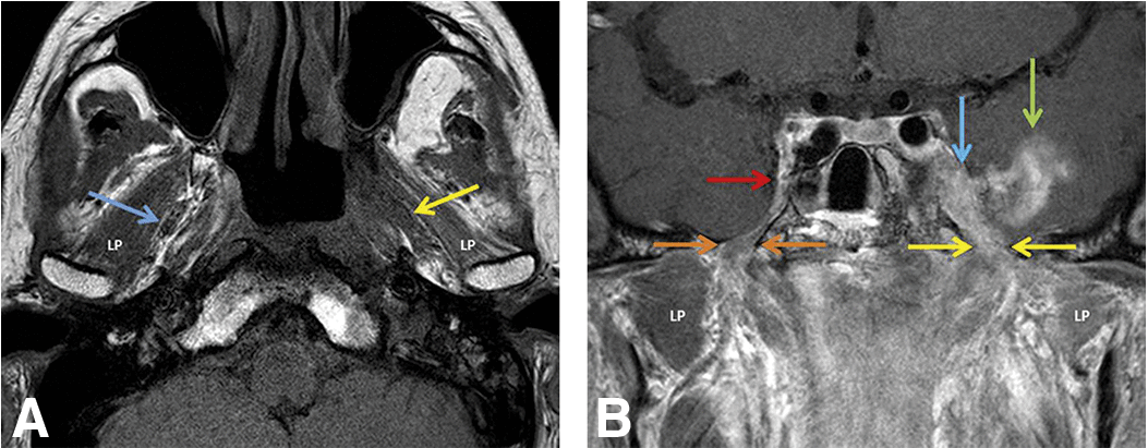
The trigeminal fat pad surrounds CN V3. Look for preservation of those straight lines in the medial masticator space between pterygoid muscles and venous plexus
Normal right fat pad and nasopharyngeal cancer spreading via the left into foramen ovale and Meckel cave:
Normal right fat pad and nasopharyngeal cancer spreading via the left into foramen ovale and Meckel cave:
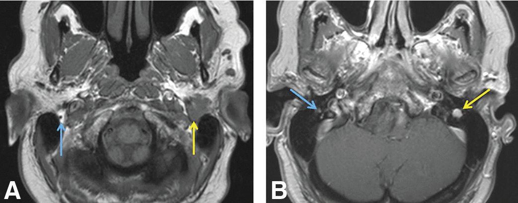
The facial nerve enters the skull at the stylomastoid foramen. There's a tiny pad of fat there!
Normal right fat pad and perineural spread of parotid mucoepidermoid cancer on the left:
Normal right fat pad and perineural spread of parotid mucoepidermoid cancer on the left:
CN V1 divides into frontal and then supraorbital nerve, which travels along the orbital roof and out the supraorbital foramen, which is nicely outlined by a bit of fat!
Right normal fat and left perineural spread of skin cancer from forehead:
Right normal fat and left perineural spread of skin cancer from forehead:
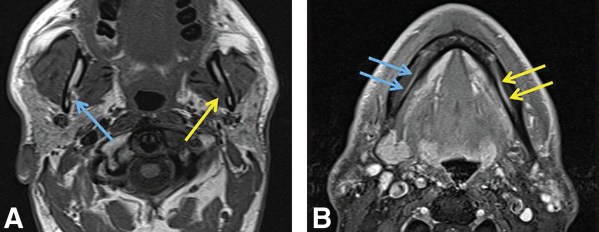
CN V3 branches into the inferior alveolar nerve, which enters the mandibular canal at the mandibular foramen, which is outlined by fat!
Right normal fat pad vs. left with perineural spread of squamous cell carcinoma from retromolar trigone
Right normal fat pad vs. left with perineural spread of squamous cell carcinoma from retromolar trigone
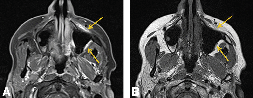
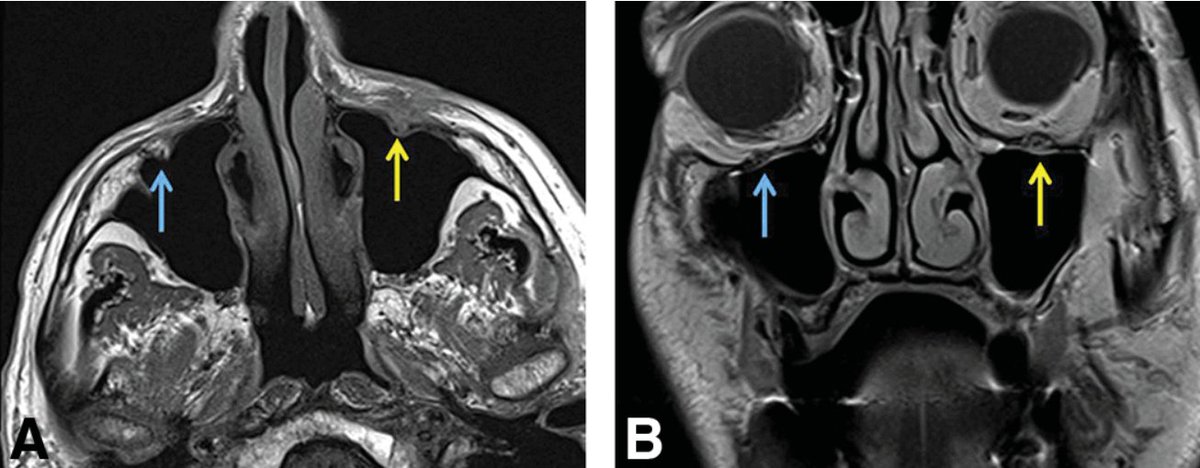
CN V2 branches into the infraorbital nerve, which exits the maxilla at the infraorbital foramen, which has a little bit of very useful fat! Also look for widening of the infraorbital canal.
Right normal foramen vs. left perineural spread from skin cancer on the cheek:
Right normal foramen vs. left perineural spread from skin cancer on the cheek:
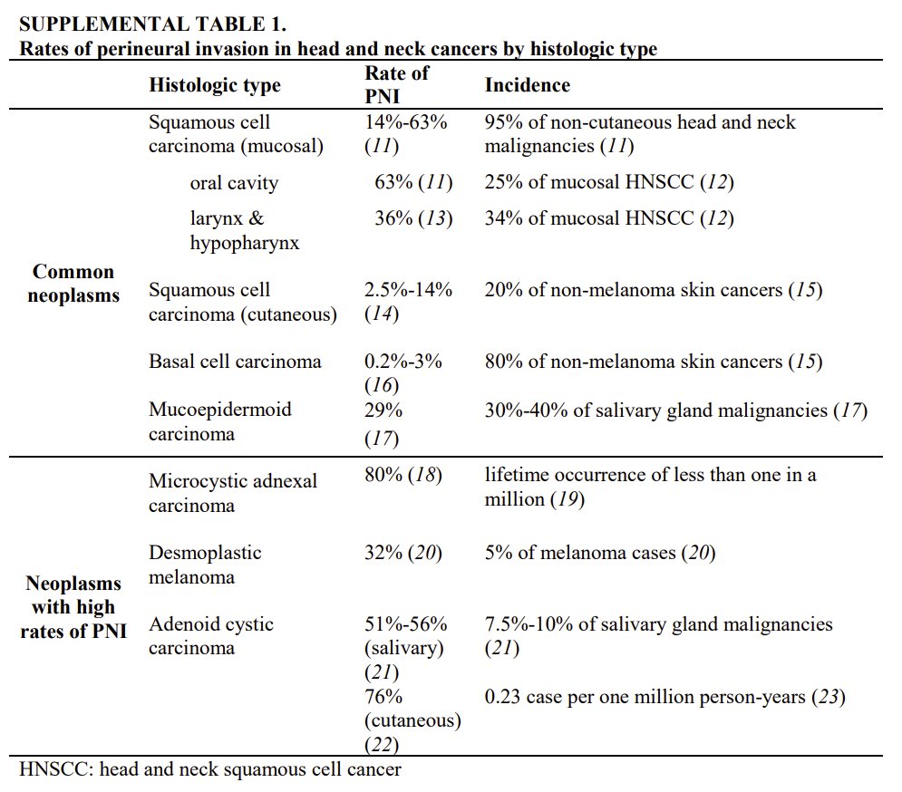
The most common histology of head and neck cancer with perineural spread is squamous cell carcinoma, because it's so common, but mucoepidermoid and adenoid cystic carcinoma are also high risk offenders
Tongue base cancer can spread along CN XII (hypoglossal)
https://pubs.rsna.org/doi/pdf/10.1148/rg.336135501
https://pubs.rsna.org/doi/pdf/10.1148/rg.336135501
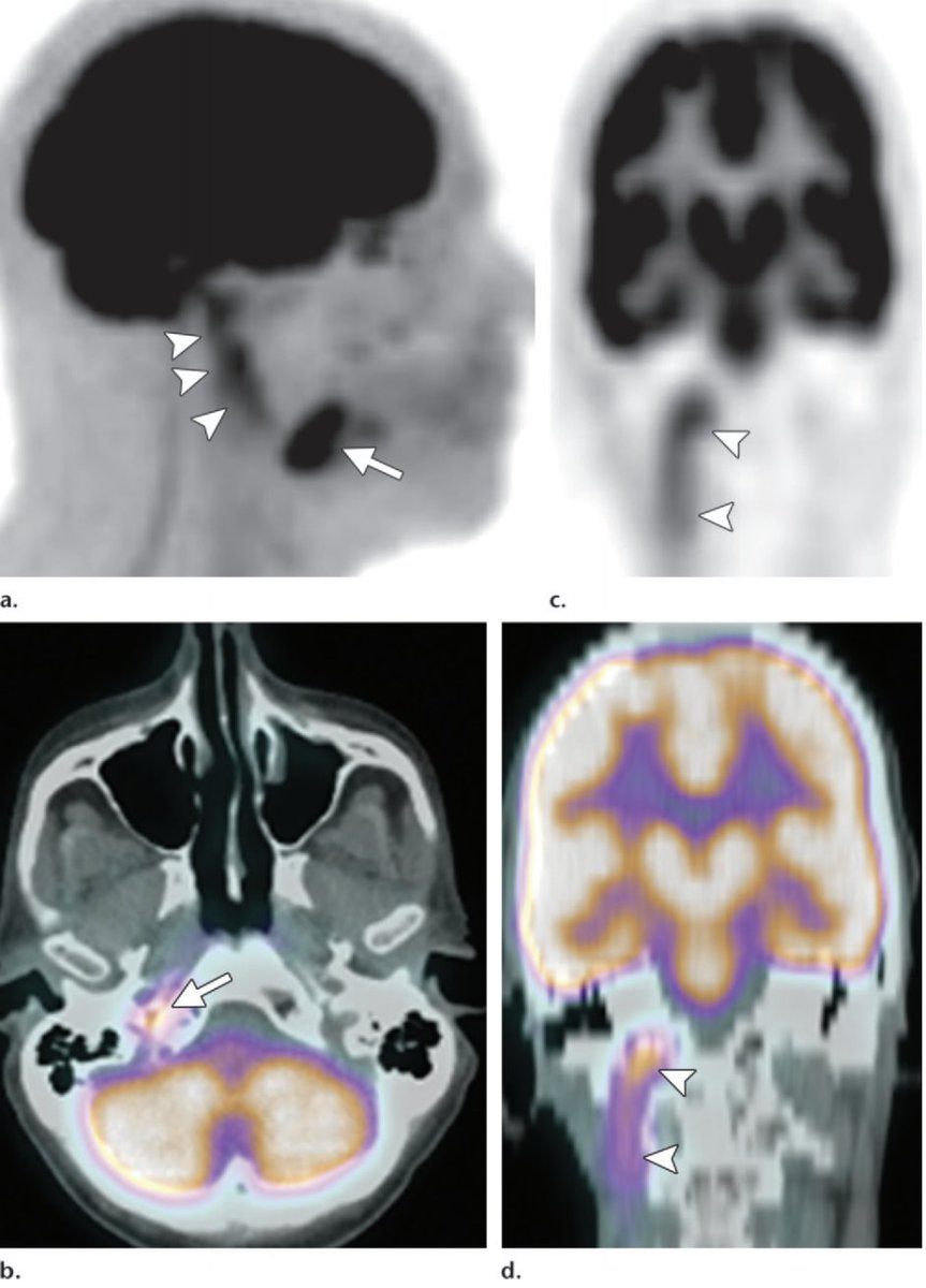
Hemitongue denervation changes (edema, enhancement, and/or fatty change, straight border down the midline) are an important clue to involvement of the hypoglossal nerve
Nasopharyngeal carcinoma can directly extend to the skull base including the hypoglossal canal. These cases won't be subtle - you don't really need a fat pad to help you here.
http://doi.org/10.1007/s003300050492
http://doi.org/10.1007/s003300050492
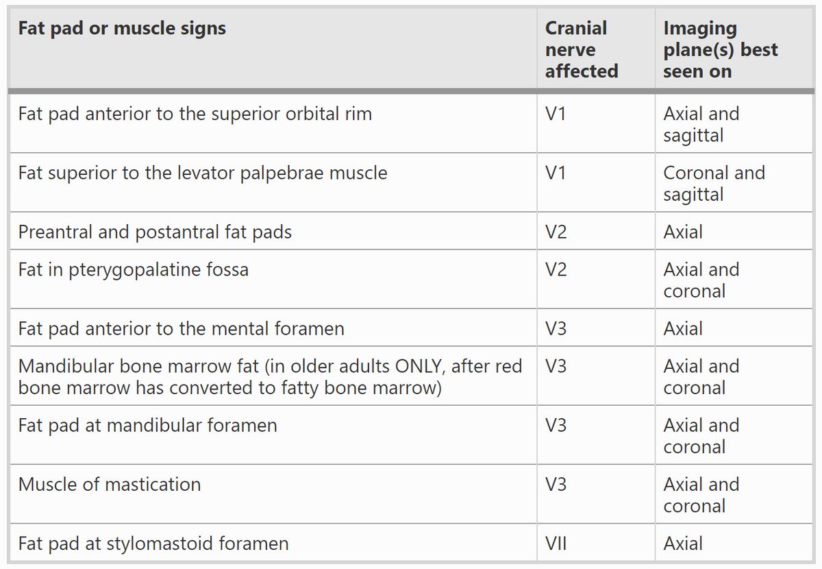
Here's a nifty table of clinical presentation, imaging landmarks, and secondary imaging signs of perineural tumor spread along cranial nerves
https://jnm.snmjournals.org/content/60/3/304.short
https://jnm.snmjournals.org/content/60/3/304.short
That's all folks.
Actually, bonus fact:
The auriculotemporal nerve is a potential connection between CN V3 and VII for perineural spread. Look just posterior to the mandible!
http://ajnr.org/content/23/2/303
Normal on the right vs. mucoepidermoid carcinoma from left parotid
Actually, bonus fact:
The auriculotemporal nerve is a potential connection between CN V3 and VII for perineural spread. Look just posterior to the mandible!
http://ajnr.org/content/23/2/303
Normal on the right vs. mucoepidermoid carcinoma from left parotid
(There are actually many potential connections between facial and trigeminal nerves, but maybe we talk mainly about the auriculotemporal because it is the biggest or most proximal? https://doi.org/10.1016/j.jpra.2017.01.005)

 Read on Twitter
