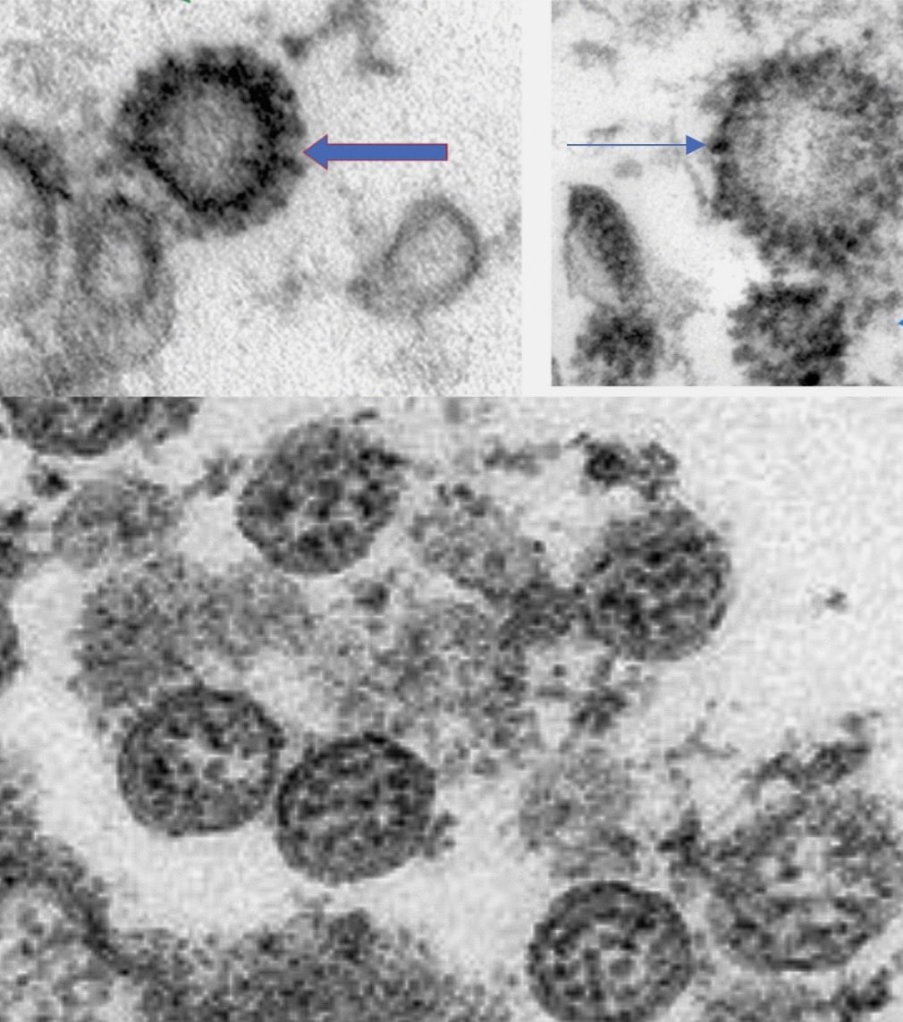
A new publication states that the top two images are Sars2 in testis. Recall that clathrin pits look like Coronaviruses, and can be an easy trap, but they can be differentiated by the cross linked structures between the spike-like structures. Left arrow.
The right top image is more curious. It looks more like dilatated rough endoplasmic reticulum than anything else, the arrow points to a ribosome, but leydig cells have all sorts of bizarre ultrastructural aggregations. It is not corona virus tho.
The bottom image is of SARS-Cov-2 particles emerging from a cell. Many of them do not have well developed spikes visible, but they do possess strikingly visible nucleocapsids inside the particles. #pathtwitter @DrGeeONE @ariella8 @TheKarenPinto
Electron microscopy can be a powerful technique when used correctly. It is disturbing that more journals do not have EM experts among reviewers to screen electron micrographs before publication. It is something that the field of pathology needs to address.

 Read on Twitter


