Is there a #POCUS method to predict weaning and extubation success? See the case and #tweetorial below.
@viamdinh @MprizzleER @iceman_ex @nickmmark @ACEP_EUS @SCCM @SAEMAEUS @SCUFellowships @UCLAEMRes
@viamdinh @MprizzleER @iceman_ex @nickmmark @ACEP_EUS @SCCM @SAEMAEUS @SCUFellowships @UCLAEMRes
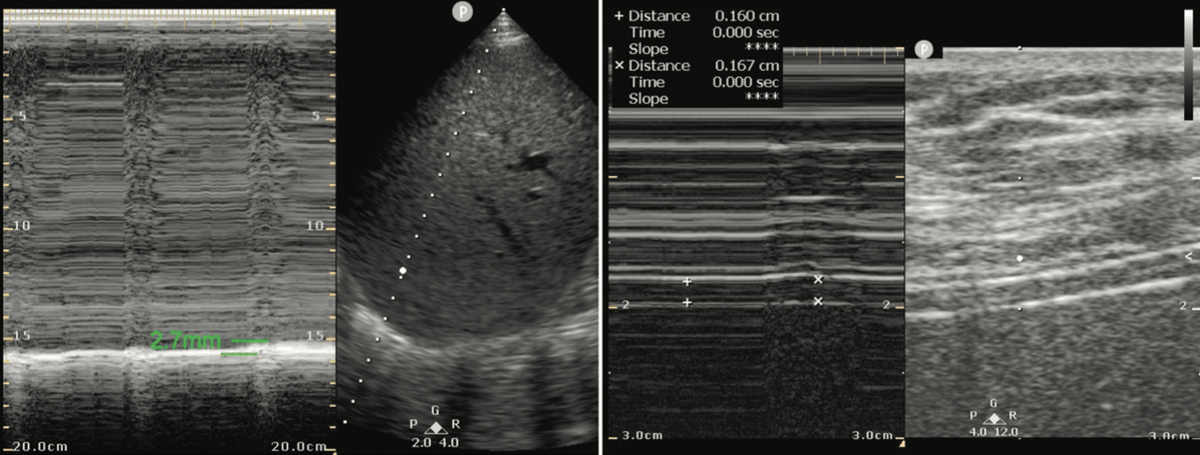
Your 89-year-old female patient is improving after her initial admission to the ICU for respiratory failure due to pneumonia. She is alert and normotensive and signals her desire to be removed from the ventilator. On her spontaneous breathing trial, you perform this thoracic US:
What is your assessment?
She is likely to fail weaning. The patient has evidence of diaphragmatic dysfunction (DD) on thoracic ultrasound. In a recent meta-analysis, this was associated with a sensitivity of 85% and 8.8 odds ratio of spontaneous breathing trial failure as opposed to those without DD.
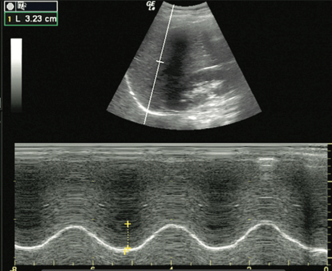
DD is assessed by measuring the diaphragmatic excursion (DE) and the diaphragmatic thickening fraction (DTF). [*eds: not our abbreviation]. For DE, place a low-frequency transducer in the mid to post-axillary line with the indicator pointed cephalad, similar to a FAST window.
Place the M-mode line at the juncture of the diaphragm and vertebral column or the costal-diaphragmatic junction. The tracing will reveal peaks with the amplitude of the peaks directly related to the inferior displacement of the diaphragm.
A DE value of ≤10-15 mm for normal spontaneous breathing and <25 mm for maximal inspiratory effort is considered abnormal. The figure below shows normal diaphragmatic excursion using M-mode.
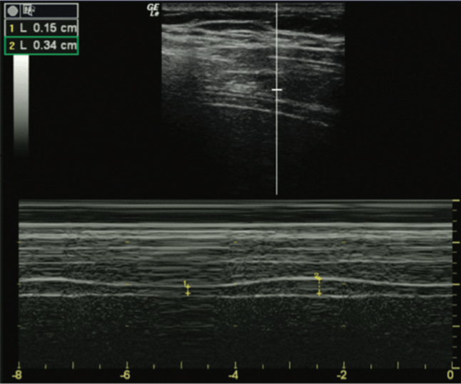
For DTF measurement, a high-frequency transducer is used in the same position as DE measurement, and M-mode is also employed. The diaphragm thickness is less during expiration (DTe) and greater than inspiration (DTi). The DTF is calculated as (DTi – DTe)/DTe.
A diaphragmatic thickening fraction <30% (with a range of 24%–35% in most studies) is considered abnormal. The figure below shows normal diaphragmatic thickening fraction using M-mode.
Our patient had a DTF of 4.4% and DE of 2.7 mm, so there is clear diaphragmatic dysfunction.
Learning Point: A DE value of ≤10-15 mm for normal spontaneous breathing and <25 mm for maximal inspiratory effort, or DTF < 30%, is associated with diaphragmatic dysfunction.
Learning Point: A DE value of ≤10-15 mm for normal spontaneous breathing and <25 mm for maximal inspiratory effort, or DTF < 30%, is associated with diaphragmatic dysfunction.
Key references:
Kim WY, Suh HJ, Hong SB, Koh Y, Lim CM. Diaphragm dysfunction assessed by US: influence on weaning from mechanical ventilation. Crit Care Med. 2011;39(12):2627–2630.
Kim WY, Suh HJ, Hong SB, Koh Y, Lim CM. Diaphragm dysfunction assessed by US: influence on weaning from mechanical ventilation. Crit Care Med. 2011;39(12):2627–2630.
Zambon M, Greco M, Bocchino S, Cabrini L, Beccaria PF, Zangrillo A. Assessment of diaphragmatic dysfunction in the critically ill patient with ultrasound: a systematic review. Intensive Care Med. 2017;43(1):29–38.
Thanks to our Resuscitative US chapter contributors, @IZBarjaktarevic and team, for this case! For more information on the 1st POCUS review book:
https://www.pocus101.com/the-ultrasound-board-review-book-is-out-exclusive-pocus-101-discount/
https://www.pocus101.com/the-ultrasound-board-review-book-is-out-exclusive-pocus-101-discount/

 Read on Twitter


