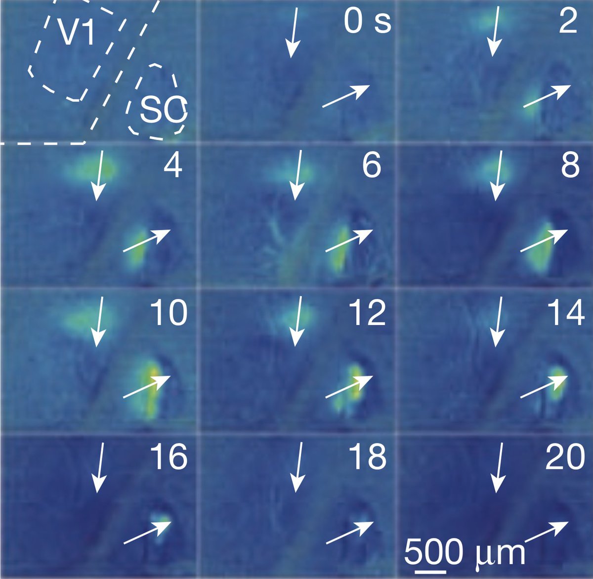
Day 2 of my Twitter #thesis. Today's topic: How spontaneous waves of activity shape the developing brain
(Figure from Ackman et al 2012 https://doi.org/10.1038/nature11529)
(Figure from Ackman et al 2012 https://doi.org/10.1038/nature11529)
Again, it all dates back to Hubel and Wiesel. Retinotopic organization is present even before an animal has had any visual experience https://doi.org/10.1113/jphysiol.1970.sp009022
Retinotopic maps are loosely established early in embryological development by genetically programmed gradients of molecular guidance cues. There's a really nice (and brief) review of that process in this 2012 review by Espinosa and Stryker https://dx.doi.org/10.1016%2Fj.neuron.2012.06.009
Later in prenatal development (or early postnatal development in mice/ferrets), retintopy is progressively refined by waves of spontaneous activity that originate in the retina and propagate throughout the developing visual system https://doi.org/10.1126/science.2035024
Blocking retinal waves (by pharmacologically or genetically disrupting retinal ACh signaling) leads to disordered retinotopy in the adult https://doi.org/10.1016/j.neuron.2005.09.015
Similar waves of activity occur in the spinal cord, where they trigger spontaneous embryological movements thought to promote self-organization of pattern-generating circuits in the spinal cord https://doi.org/10.1038/nature01719
Peripheral waves are relayed to the developing cortex and amplified by recurrent thalamocortical excitation, producing a characteristic spindle-burst electrical pattern thought to promote activity-dependent plasticity https://doi.org/10.1038/nature03132
Similar waves are generated in other developing structures, like the cochlea, cerebellum, hippocampus and neocortex. They are generated by a variety of mechanisms but they are all thought to promote self-organization of the CNS https://doi.org/10.1038/nrn2759

 Read on Twitter


