#MedTwitter #EmergencyMedicine #USMLE
 Thread: intracranial hemorrhages
 Frequently asked questions
( if not always )
)
cont..
 Thread: intracranial hemorrhages
 Frequently asked questions
 ( if not always
 )
) cont..
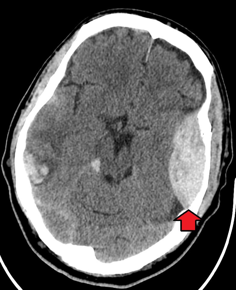
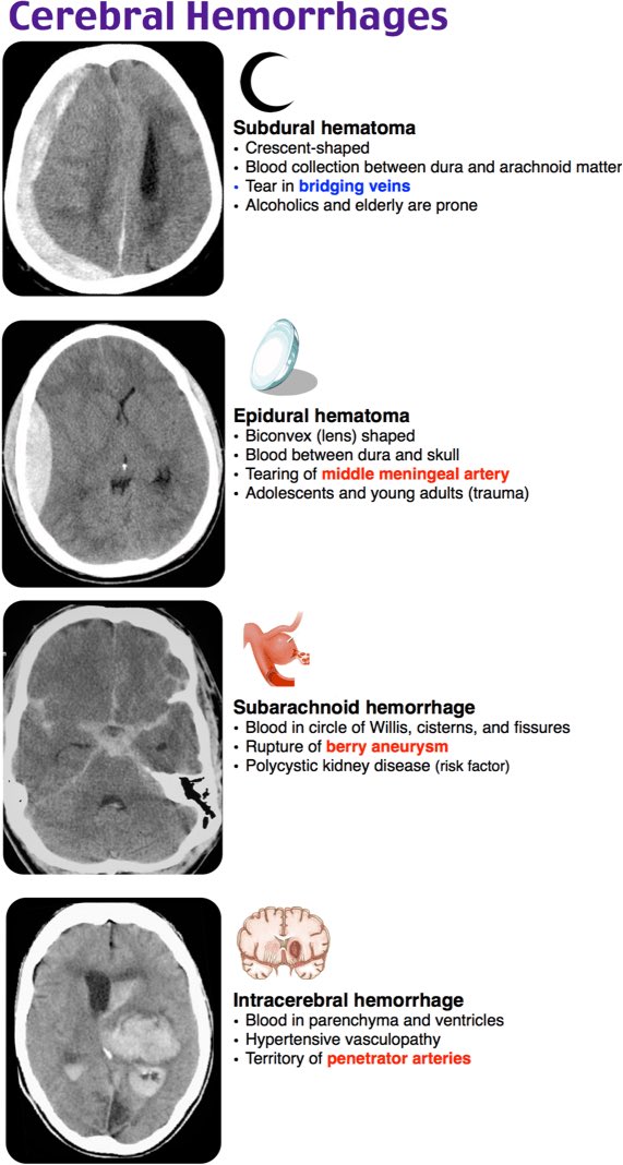
1- epidural hemorrhages ( EDH)
- Caused by laceration of meningeal arteries ( middle )
- Sometimes Associated with skull fractures ( temporal bone )
- Lenticular ( biconvex ) hematomas on CT scan.
- Caused by laceration of meningeal arteries ( middle )
- Sometimes Associated with skull fractures ( temporal bone )
- Lenticular ( biconvex ) hematomas on CT scan.
Sign and symptoms:
 +/- initial loss of consciousness
 50% have lucid interval for several hours followed by obtundation ( patient recovers then deteriorate)
 Nausa and vomiting, loss of consciousness and seizures
 +/- initial loss of consciousness
  50% have lucid interval for several hours followed by obtundation ( patient recovers then deteriorate)
 Nausa and vomiting, loss of consciousness and seizures
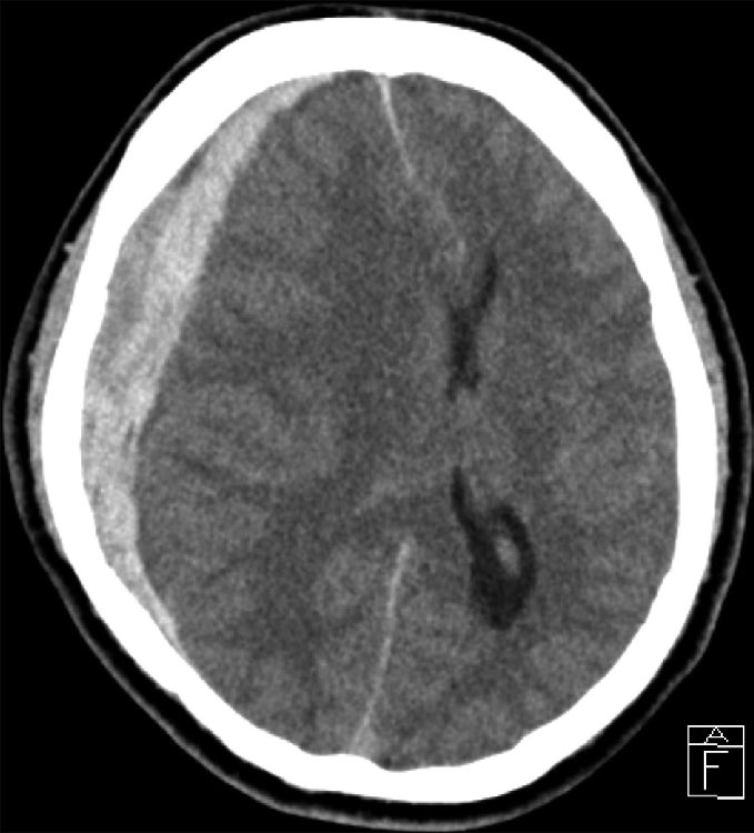
2- Subdural hemorrhage 
- Caused by laceration of the bridging corrical veins.
- More common in elderly 2ry to brain atrophy.
- Vomiting, weakness, confusion, headache, memory loss, speech disturbance and seizure.
- Crescentic shape in CT scan

- Caused by laceration of the bridging corrical veins.
- More common in elderly 2ry to brain atrophy.
- Vomiting, weakness, confusion, headache, memory loss, speech disturbance and seizure.
- Crescentic shape in CT scan
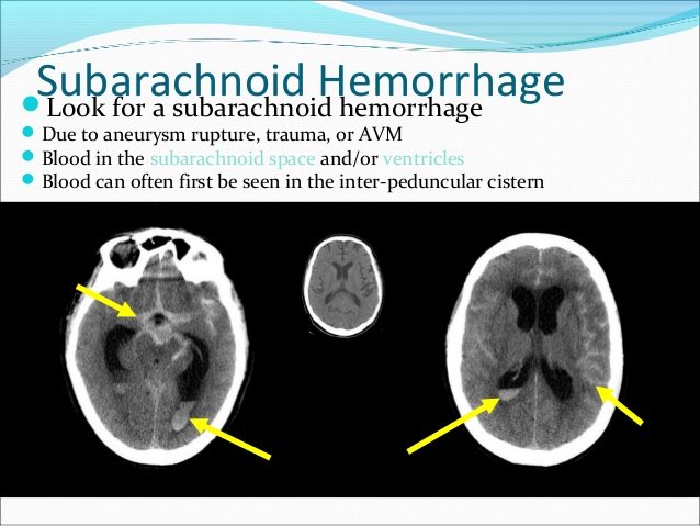
3- Subaracnoid hemorrhage ( SAH )
- Caused by ruptured aneurysm, arteriovenous malformation, direct injury to the pia vessels or extention from intraventricular hemorrhage.
- Nausa and vomiting, confusion or obtundation.
- Caused by ruptured aneurysm, arteriovenous malformation, direct injury to the pia vessels or extention from intraventricular hemorrhage.
- Nausa and vomiting, confusion or obtundation.
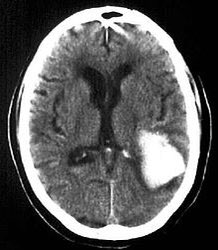
4- intraparenchymal hemorrhage
- caused by laceration of cortical parenchyma due to Contusion (coup or countercoup)
- patient may have loss of consciousness and seizures
- depends on anatomical location
- caused by laceration of cortical parenchyma due to Contusion (coup or countercoup)
- patient may have loss of consciousness and seizures
- depends on anatomical location

 Read on Twitter
