#Tweetorial below: I read a #whyCMR with @Talalkfo and @chen_alvin_s recently in which we identified a severely dilated LV in a patient with severe MR. The referring emailed me asking why the #EchoFirst only had a diameter of 5.7 cm. "Which one is right?"
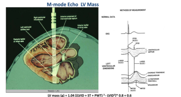
Per @ASE360 convention, dating to the time when M-mode #EchoFirst was frequently used for LV linear dimensions and mass, LVEDD is measured < 1 cm within the tips of the MV. ( https://bit.ly/38K9DM9 )
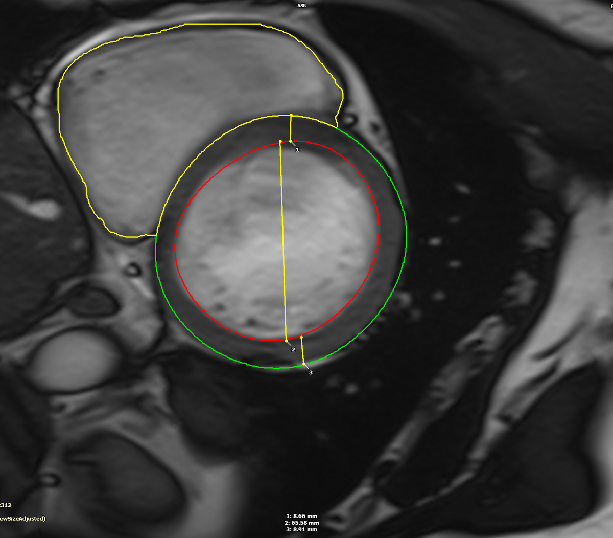
The convention in #whyCMR is to measure the LV linear dimensions in the slice distal to the MV as shown below which was 6.7 cm, consistent with the patient's LV volumes which were the severe range.
I went back and measured the LV dimension on #EchoFirst just distal to the basal septum. Lo and behold, the LV measured 6.7 cm.
This reminded me of a paper @JournalASEcho ( https://bit.ly/2BQTXei ) from @mikepicardmd @FrailtyMD + colleagues that showed measurement in the mid LV produces more accurate estimates of LV mass, size, and function c/w #whyCMR.
This becomes especially relevant in those with basal septal hypertrophy, which is very common (~19%) in the patients we see ( https://bit.ly/3fl4ay6 ), increases with age, and is in part due to more acute angulation of the aorta vs. the LV major axis.
So what to do when surgical cutoffs for intervention etc. are based on traditional #EchoFirst measurements? Should the guidelines change to reflect measurement more distal in the LV? What do others report in their #EchoFirst lab? Fin. @onco_cardiology @purviparwani @LucySafi

 Read on Twitter


