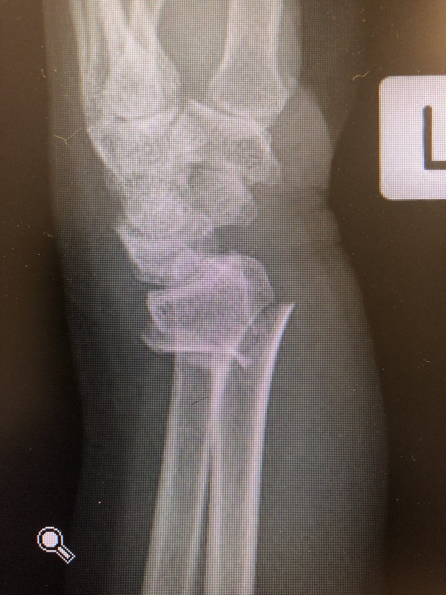
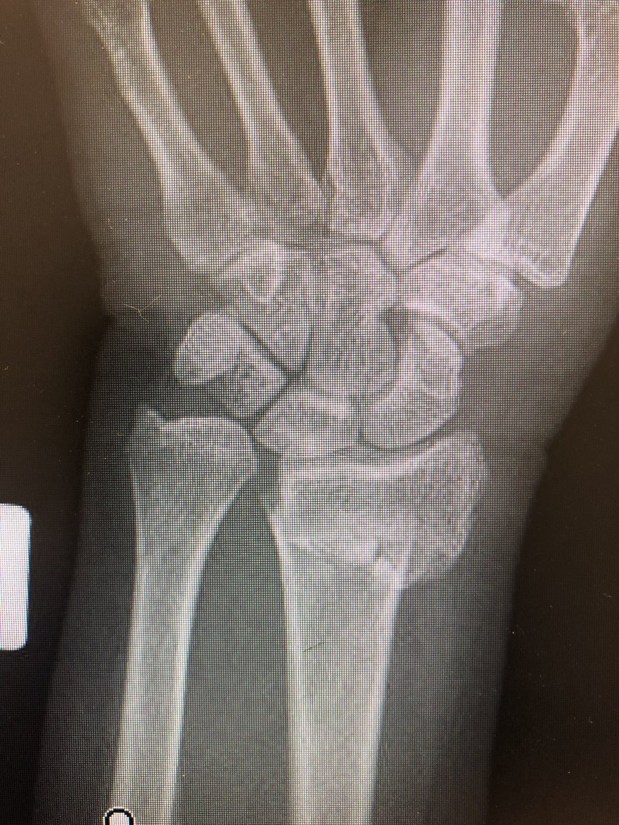
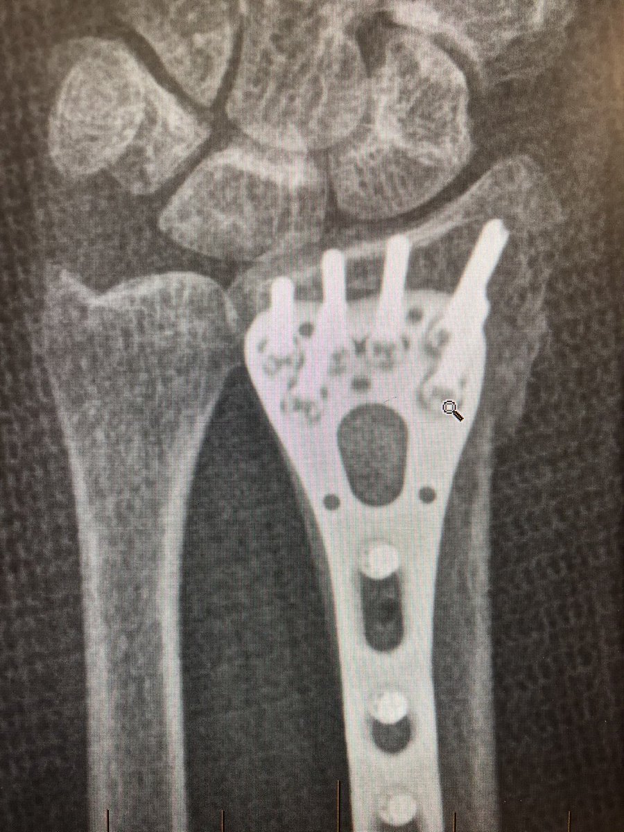
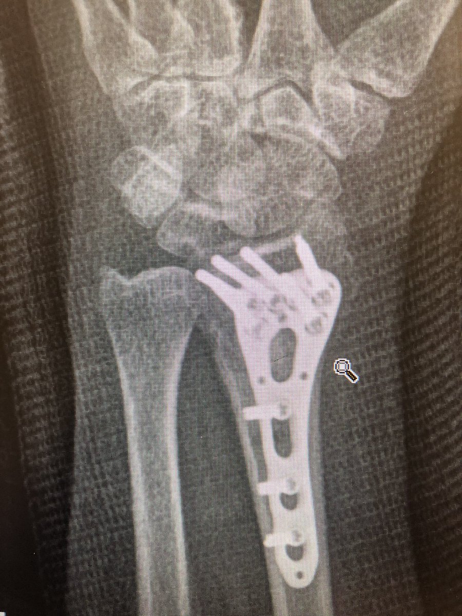
Simple everyday distal radius fracture. 60 yr old nurse. Medically well. #OrthoTwitter
I see conventionally this would be called an ‘extra-articular’ fracture.
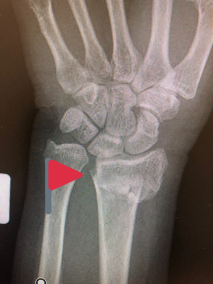
Do we oversimplify this? Isn’t DRUJ a ‘joint’?
Isn’t the distal ulna getting all into the business of the carpals a ‘joint disruption’?
I would fix this.
Do we oversimplify this? Isn’t DRUJ a ‘joint’?
Isn’t the distal ulna getting all into the business of the carpals a ‘joint disruption’?
I would fix this.
Point 1:
The proximal fragment radius spike has a risk of translational mal-reduction and this can bother rotation of the forearm.
Reduce this by putting a ‘lamina spreader’ in the inter-osseus space Before locking the shaft screws and clear the DRUJ.
The proximal fragment radius spike has a risk of translational mal-reduction and this can bother rotation of the forearm.
Reduce this by putting a ‘lamina spreader’ in the inter-osseus space Before locking the shaft screws and clear the DRUJ.
Point 2:
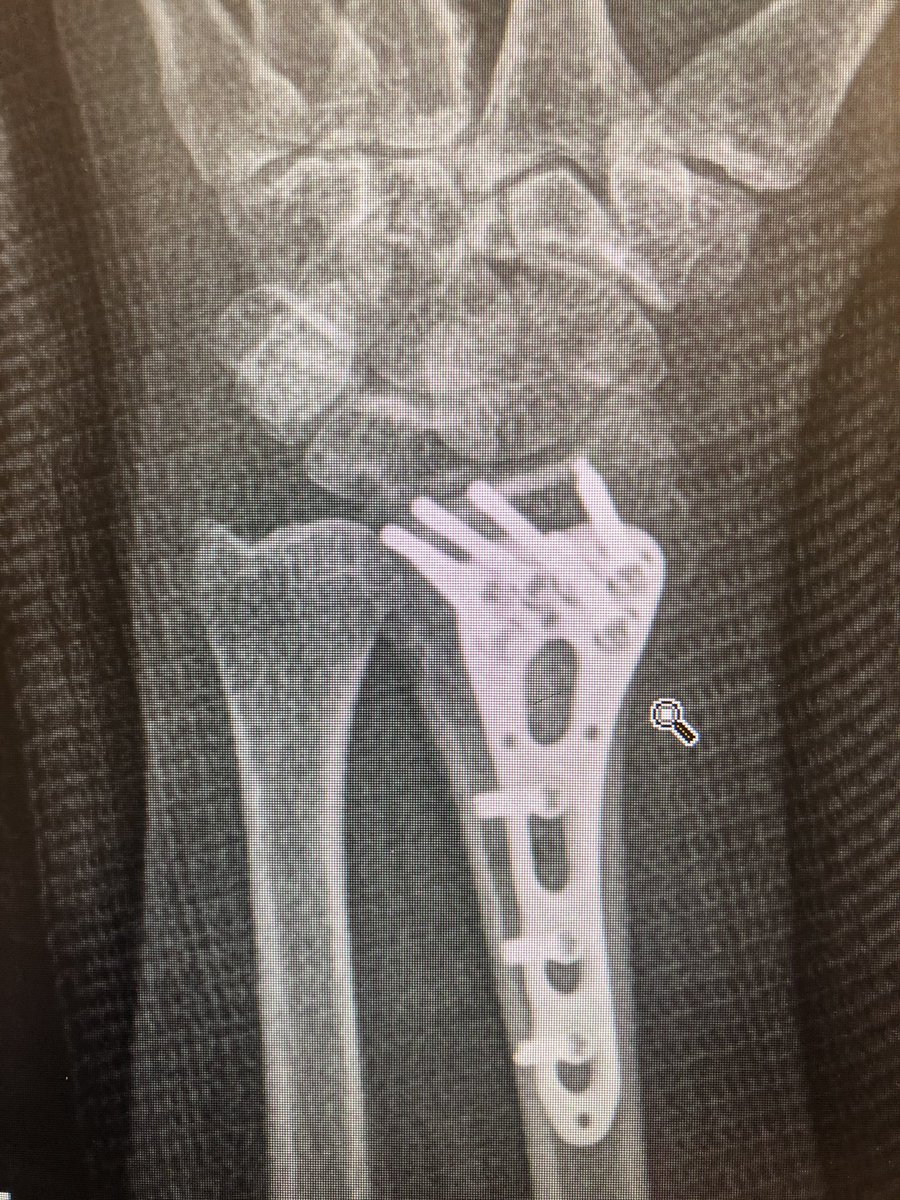
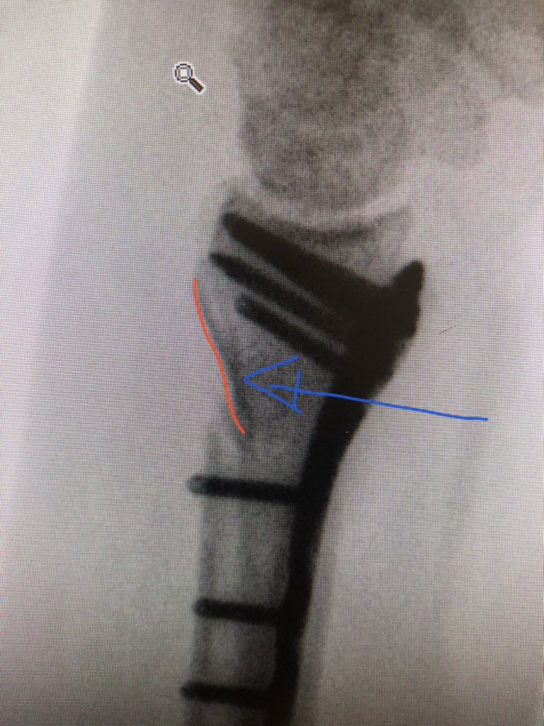
The dorsal metaphyseal comminution. The dorsal cortex fragment is ‘fallen in’ inside the osteoporotic metaphyseal void.
I addressed this after plating finished, made a 4.5 mm drill hole in the ‘window’ of the plate to push the fragment and fill in graft substitute.
The dorsal metaphyseal comminution. The dorsal cortex fragment is ‘fallen in’ inside the osteoporotic metaphyseal void.
I addressed this after plating finished, made a 4.5 mm drill hole in the ‘window’ of the plate to push the fragment and fill in graft substitute.
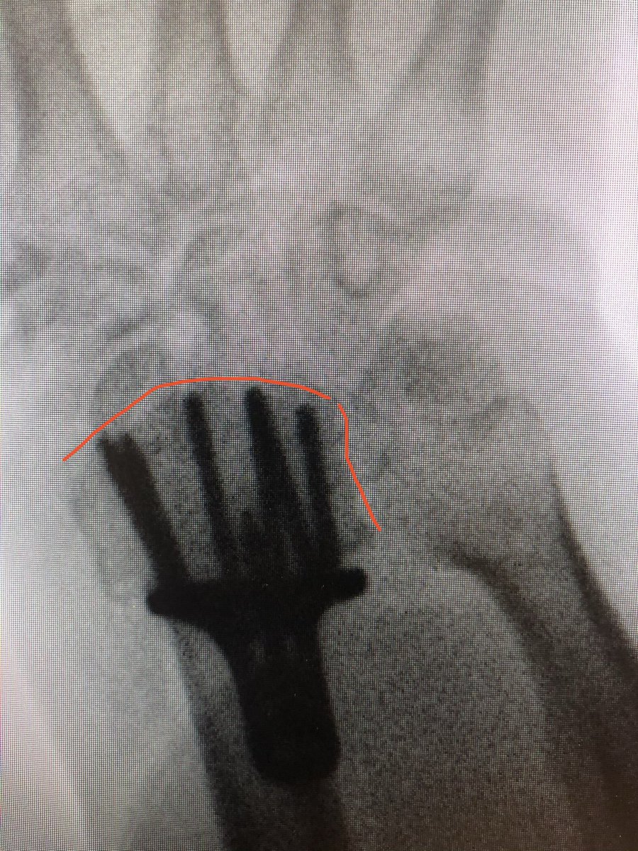
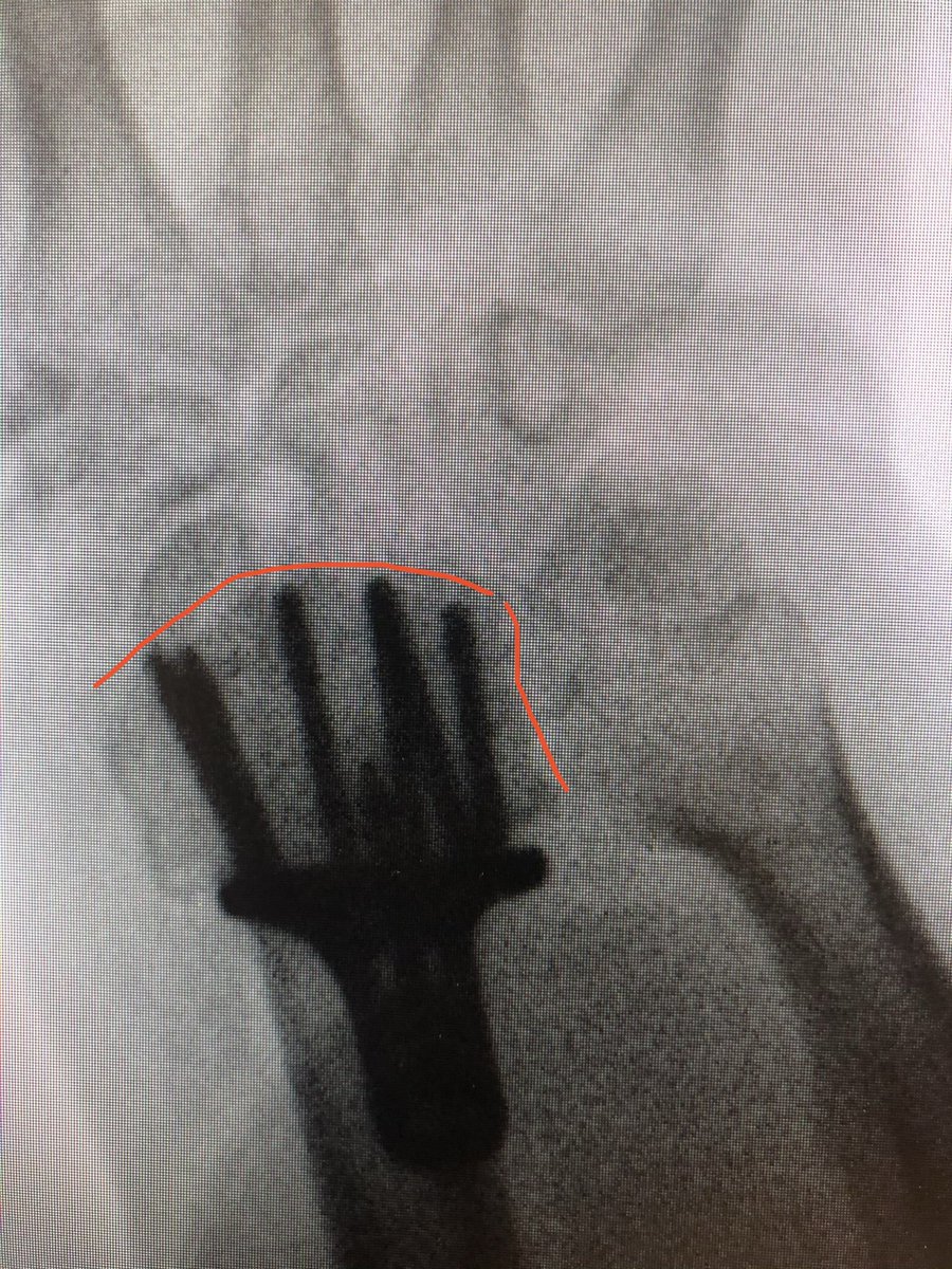
Point 3: the tangential view.
The wrist flexed ‘dorsal tangential view’ or as we locally call it ‘Lleyton Hewitt View’ is increasingly popular (published from our institutes with my colleagues too), but I find the ‘extended tangential’ or ‘DRUJ View’ more informative.
The wrist flexed ‘dorsal tangential view’ or as we locally call it ‘Lleyton Hewitt View’ is increasingly popular (published from our institutes with my colleagues too), but I find the ‘extended tangential’ or ‘DRUJ View’ more informative.
Here’s one of the works my department has done in this area. https://dspace.flinders.edu.au/xmlui/bitstream/handle/2328/38390/Bergsma_Volar_AM2018.pdf?sequence=1&isAllowed=y
As compared to the dorsal tangential view, this DRUJ view gives better light contrast to view the dorsal rim and also additional information about the DRUJ to make sure there is no screw protrusion into the ulnar notch.
This was the patient at 2 weeks. Already on range of motion. The flat lateral X-ray always looks like the screws are in the joint. That’s because of the distal radius joint inclination angle.
Take intra-operative 20 degree elevated lateral shot to see through the joint.
Take intra-operative 20 degree elevated lateral shot to see through the joint.

 Read on Twitter

