1. “STEMI equivalent” #ECG patterns is crucial for every
 dealing with #ACS.
-in 10-25% pts for urgent #PCI
 Wellens’ syndrome
 de Winter sign
 hyperacute T waves,
 left bundle branch block (LBBB)
 right bundle branch block (RBBB)
 https://www.ajconline.org/article/S0002-9149(19)30827-6/fulltext?mobileUi=0

 dealing with #ACS.
-in 10-25% pts for urgent #PCI
 Wellens’ syndrome
  de Winter sign
 hyperacute T waves,
  left bundle branch block (LBBB)
 right bundle branch block (RBBB)
  https://www.ajconline.org/article/S0002-9149(19)30827-6/fulltext?mobileUi=0
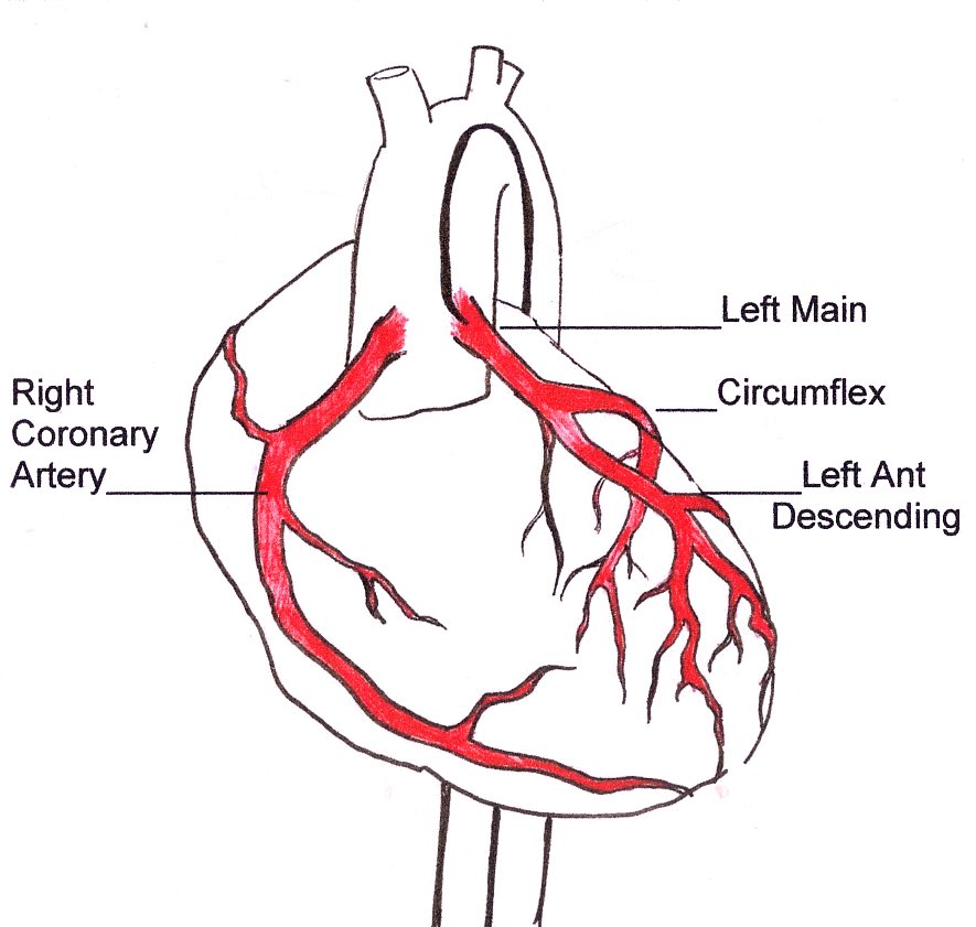
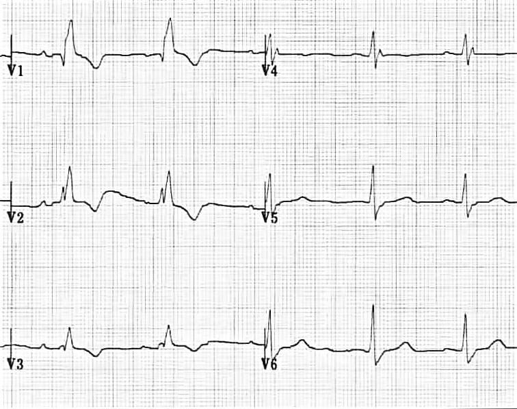
2. Wellen’s syndrome
(other names Wellens' sign, Wellens' warning, Wellens' waves):
is a pattern of deeply inverted or biphasic T waves in V2-3, which is highly specific for a critical stenosis of the left anterior descending artery (LAD)
 Wellen’s syndrome
(other names Wellens' sign, Wellens' warning, Wellens' waves):
is a pattern of deeply inverted or biphasic T waves in V2-3, which is highly specific for a critical stenosis of the left anterior descending artery (LAD)
3. de Winter sign
-ECG abnormality described by de Winter et al. in 1998
-Characterized by 1-3 mm of ST-depression with upright, symmetrical T-waves
-Suspicious for proximal occlusion of the LAD
-Recognized as a STEMI equivalent by Rokos et al. in 2010
 de Winter sign
-ECG abnormality described by de Winter et al. in 1998
-Characterized by 1-3 mm of ST-depression with upright, symmetrical T-waves
-Suspicious for proximal occlusion of the LAD
-Recognized as a STEMI equivalent by Rokos et al. in 2010
4. Hyperacute T waves
Broad, asymmetrically peaked or ‘hyperacute’ T-waves are seen in the early stages of ST-elevation MI (STEMI) and often precede the appearance of ST elevation and Q waves.
They are also seen with Prinzmetal angina
 Hyperacute T waves
Broad, asymmetrically peaked or ‘hyperacute’ T-waves are seen in the early stages of ST-elevation MI (STEMI) and often precede the appearance of ST elevation and Q waves.
They are also seen with Prinzmetal angina
5. Left bundle branch block
QRS duration of>120 ms
Dominant S wave in V1
Broad monophasic R wave in lateral leads(I,aVL,V5-V6)
Absence of Q waves in lateral leads (I, V5-V6; small Q waves are still allowed in aVL)
Prolonged R wave peak time>60ms in left precordial leads(V5-6)
 Left bundle branch block
QRS duration of>120 ms
Dominant S wave in V1
Broad monophasic R wave in lateral leads(I,aVL,V5-V6)
Absence of Q waves in lateral leads (I, V5-V6; small Q waves are still allowed in aVL)
Prolonged R wave peak time>60ms in left precordial leads(V5-6)

 Read on Twitter


